| Join US on Facebook.com/Amygdala-PanicDisorder - - - - - - (C) 2015/2016 OUR THANKS TO PUBMED.GOV |

|
|
|
|
 |
| Whats a Panic Attack? New forms of Amygdalectomy: less tissue, less risks Scientific discoveries |
| Home Institutional Who's who Definitions Epidemology Patient's rights |
|
Scientific discoveries - Amygdala, the brain fear/worries/stress center: examination of 2000 studies - (on 74.500 total, www.PubMed.gov) ANATOMY AND FUNCTIONALIY OF THE CENTRAL AMYGDALA The Amygdala is responsible for detecting and responding to threats [..] For example, the Amygdala outputs alter information processing in diverse regions of the brain. One important set of outputs result in the secretion of chemicals throughout the brain (norepinephrine, acetylcholine, dopamine, serotonin) and body (hormones such as adrenalin and cortisol). In situations of threat/danger, these chemicals alert the organism that something important is happening. As a result attention systems in the neocortex guide a perceptual search inside the environment for an explanation of the highly aroused state. 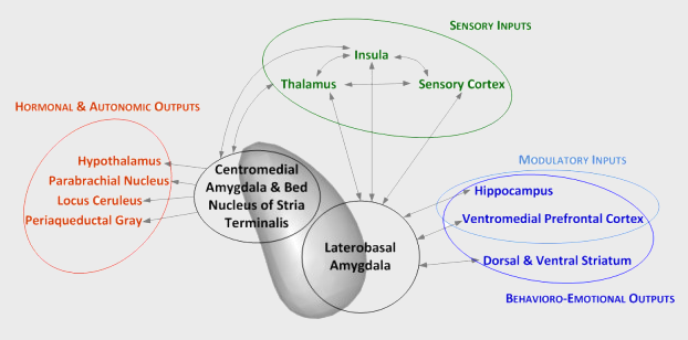 * Microcircuits Controlling Learned Fear / 2014 - Sevil Duvarci1 and Denis Pare, Neuron vol.82 Lateral amygdala is main point of entry for sensory inputs, whereas Central amigdala is the main and source of projections that output to brainstem fear effector structures. * The role of the Amygdala in fear and anxiety / 1992 - Annual Review of Neuroscience Amygdala is the primary structure of the brain responsible for Fight or Flight response (FF). [..] The amygdala is directly associated with Conditioned Fear. Conditioned Fear is the framework used to explain the behavior produced when an originally neutral stimulus is consistently paired with a stimulus that evokes fear. The amygdala represents a core fear system in the human body, which is involved in the expression of conditioned fear. Fear is measured by changes in autonomic activity including increased heart rate, increased blood pressure, as well as in simple reflexes such as flinching or blinking. The central nucleus of the amygdala (CeA) has direct correlations to the hypothalamus and brainstem areas directly related to fear and anxiety. This connection is evident from studies of animals that have undergone amygdalae removal. Such studies suggest that animals lacking an amygdala have less fear expression and indulge in non-species-like behavior. Mammals have very similar ways of processing and responding to danger (rodents, monkeys, humans). By observing the amygdalas functions, people can determine why one rodent may be much more anxious than another. There is a direct relationship between the activation of the amygdala and the level of anxiety the subject feels. Feelings of anxiety start with a catalyst an environmental stimulus that provokes stress. This can include various smells, sights, and internal feelings that result in anxiety. The amygdala reacts to this stimuli by preparing to either stand and fight or to turn and run. This response is triggered by the release of adrenaline into the bloodstream. Consequently, blood sugar rises, becoming immediately available to the muscles for quick energy. * Amygdala. Afferent and Efferent Connectivity. / 2009 - Baylor University Waco, Texas CeA lesion and stimulation studies have shown cortical, hypothalamic, and brain stem regions to be target areas (Iwata et all.1987; LeDoux, Iwata, Cicchetti, & Reis, 1988; LeDoux, 2000; Sah et al., 2003; Turner, Mishkin, & Knapp, 1980 ;Walker & Davis, 2002). CeA efferents modulate specific behavioral and autonomic responses to fear, anxiety, and stress (Davis, 1997; Rosen & Schulken, 1998; Sah et al., 2003). CeAs connection to the hypothalamus allows activation of the sympathetic nervous system (HPA, skin response, pupil dilation,.. in response to fear). * The hypothalamus-pituitary-adrenal axis / 2008 - Besedovsky, Hugo; Chrousos, George; Rey, Adriana Del; ISBN 9780444530400 HPA axis is a set of interactions among three endocrine glands: hypothalamus, pituitary gland (below hypothalamus) and adrenal glands (on top of the kidneys). HPA axis, as major part of the neuroendocrine system, controls reactions to stress (ie aggressivity/fear fight/flight response) and regulates many body processes through hormones like noradrenaline, adrenaline and cortisol. Anatomical connections between Amygdala and Hypothalamus facilitate activation of the HPA axis: sensory information arriving at the lateral aspect of the amygdala (LA) is processed and conveyed to the central nucleus (CeA), which projects to several parts of the brain involved in responses to fear. At the hypothalamus, fear-signaling impulses from CeA activate both the sympathetic nervous system and the modulating systems of the HPA axis. 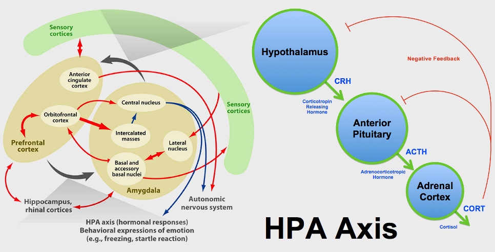
* Amygdala Microcircuits / 2012 - Denis Pare et al., Center for Molecular & Behavioral Neuroscience New Jersey Central Amygdala (fear effector) project to brainstem and hypothalamic, triggering conditioned fear responses. * Amygdala: vigilance and emotion / 2001 - M Davis, PJ Whalen, Emory U.School of Medicine, GA Big difference between basolateral amygdala (BLA) and surrounding nuclei (central, medial, cortical) in terms of cell shape, cell content and projection patterns. The BLA is a sort of switching center, an example: 1) A light paired with food can serve as a positive reinforcer by changing neural transmission in BLA which sends signals to the striatum, leading to approach behavior; 2) A light paired with shock may change neural transmission in BLA which projects to the CeA to produce the somatic, autonomic and endocrine signs of fear. * A circuit mechanism for differentiating positive and negative associations / 2015 - Praneeth Namburi et al. The Picower Institute for Learning and Memory, Department of Brain and Cognitive Sciences, Massachusetts Institute of Technology, Cambridge BLA a switching center between fear and pleasure. Synaptic plasticity in the BasoLateral Amygdala complex (BLA) mediates the acquisition of associative memories, both positive and negative. Different populations of BLA neurons may encode fearful or rewarding associations. Here we show that BLA neurons projecting to the nucleus accumbens (NAc projectors, reward response) or the centromedial amygdala (CeM projectors, fear response) undergo opposing synaptic changes following fear or reward conditioning. We find that photostimulation of NAc projectors supports positive reinforcement while photostimulation of CeM projectors mediates negative reinforcement. Photoinhibition of CeM projectors impairs fear conditioning and enhances reward conditioning. * Emotional memory manipulated: studying Hippocampus and Amygdala / 2015 Susumu Tonegawa, RIKEN-MIT Center for Neural Circuit Genetics Memories of either emotion can be reversed in the hippocampus. But the amygdala carries only positive or negative memory and cannot interchange. (the emotional valence of events cannot be modified in the amygdala when deeply-rooted) AMYGDALA FROM FISH TO HUMAN In 1819, the anatomist Karl Burdach described an almond-shaped structure that was located in the temporal lobe of the human brain, which he accordingly named the amygdala. In 1956, Weiskrantz lesioned the amygdala, demonstrating impairments in acquiring behavioural responses to shock-predictive cues (CS), increased tameness and lessening or disappearance of previously acquired fear responses. Following these early studies, amygdala lesions in both rodents and humans revealed a strong conservation of normal body and brain function, and most notably impairment were in the recognition of fearful stimuli, and in a type of emotional learning called fear conditioning. The relative enlargement of the BLA compared with the CeA between rodents and primates might result from the substantial increase in the size of cortical regions that communicate with the BLA in primates. 
AMYGDALA LESIONS * The impact of early and late damage to the human Amygdala / 2004 P.Shaw, E.J.Lawrence, C.Radboume, et al.., Centre for Neuroscience, King's College London, UK Early amygdala lesion is disruptive to the development [..] Late damage does not affect theory of mind reasoning as the Amygdala is not needed in adult life for the performance of such abstract reasoning. * Effects of Aspiration versus Neurotoxic lesions of Amygdala on emotional responses in monkeys / 1999 - Martine Meunier, Jocelyne Bachevalier, Elisabeth A. Murray, Ludis eMa lkova, Mortimer Mishkin National Institute of Mental Health, Bethesda, MD, USA, Institut Des Sciences Cognitives, CNRS, Bron, France - Fear and aggression are markedly reduced, but they are not eliminated. This is consistent with human data, which indicate that patients with bilateral amygdalectomies show attenuated affect, but are not emotionless. * Evolutionary Neuroscience. / 2009 - John H. Kaas, Elsevier, United Kingdom, UK Lesion of Amygdala in monkey demonstrated that there is massive reduction of fear and aggression. Monkeys also become more friendly sexually and non-sexually towards other monkeys. * Handbook of Biobehavioral Approaches to Self-Regulation. / 2015 - Guido H.E. Gendolla et al. Distruction of Amygdala does not disrupt emotional learning. * Amygdala lesions do not compromise the cortical network for false-belief reasoning. / 2015 - LeDoux The amygdala is not a necessary component of the cortical network for false-belief reasoning detecting. * Neuroanatomy of Social Behaviour: An Evolutionary and Psychoanalytic Perspective. / 2011 Author RALF-PETER BEHRENDT - ed. Karnac - London CEA and BNST induce freezing via parallel projections in PAG, contextual conditioned freezing can be attenuated by lesions to this regions. Patients with PTSD show evevated cerebrospinal-fluid concentrations of CRF which may sensitize (chronic activation) CRF systems in Amygdala and BNST. * The Role of the Central Nucleus of the Amygdala in Fear and Anxiety in the Primate. / 2004 - Ned H. Kalin Bilaterally lesioned monkeys displayed significantly less fear-related behavior when exposed to a snake and less freezing behavior when confronted by a human intruder. In addition, bilaterally lesioned monkeys had decreased levels of CRF (corticotrophin-releasing factor) and both lesioned groups had decreased plasma ACTH concentrations. * Autism, Neural Basis and Treatment Possibilities / 2002 - David G. Amaral, Novartis Foundation Analysis of patients with bilateral amygdala damage: Amygdala is not a central component of the Social System. * CEA lesions attenuate stress-induced anxiety behavior in rats / 2013 Hebrew Univ., Jerusalem Lesion in the CeA induces different results in anxiety and fear-behaviors. Lesioned animals display attenuation of the stress response and of stress-induced anxiety-like behavior when compared with stressed animals with sham lesions. This attenuation was paralleled by a decrease of stress-induced corticosterone levels. These results confirm the implication of the CeA in fear conditioning behavior and unravel the relevance of this brain region in the regulation of the HPA axis activity and in the onset of anxiety behavior triggered by stress. * Explicit semantic memories (hippocampus) versus implicit fearful memories (amygdala) / various Édouard Claparede (Swiss neurologist, 1873-1940) especially studied hippocampal lesioned patients. Those people presented serious amnesia: old memories were ok, but the recent past was not remembered. Therefore, Claparède made a simple experiment. When introducing himself, he hid a pin in his hand and reached to shake the patients's hand, pricking the patient. The next day, sure enough, the patient did not remember him. But when Claparède went to shake the patient's hand, he found that the patient hesitated, recognizing a threat even if not understanding the reason. Hippocampal lesioned people felt frightened, but they didn't know why. On the contrary amygdala lesioned patients were not amnesic. They recognized the doctor, remembered the hidden pin and the pain felt previous day, but they lacked of stress and worry. These experiements have been confirmed in later studies (for example: Hippocampal lesions lead to abnormal responses to social signals and degradation of social bonds. Machado et al., 2006) * Amigdala & Hippocampus. Separate ablations in Monkeys / 1991 - Stuart Zola-Morgan, Larry R. Squire, Pablo Alvarez-Royo and Robert P. Clower, University of California, San Diego Independence of Memory Functions and Emotional Behavior. Separate Contributions of the Hippocampal Formation and the Amygdala. Amygdala lesions (results): The abnormal emotional behavior associated with (bilateral) amygdala lesions was expressed as increased reactivity to the object stimuli, specifically as an increased tendency to examine and manipulate objects. This same abnormality has been observed consistently in studies of monkeys with lesions of the amygdaloid complex (Thompson et al., 1969; Horel et al., 1975; Aggleton and Passingham, 1981). No memory impairments were reported in all experiments (memory battery): 1) Delayed non matching 2) object retention 3) concurrent discrimination. On the contrary hippocampal lesions seriously impaired memory. STIMULATING AND INHIBITING AMYGDALA The amygdala, the hub of fear processing networks, is closely associated with the pathogenesis of panic disorder (PD). Electrical or chemical stimulation of the amygdalar central nucleus causes constellation of symptoms that are very similar to those of panic attacks. Electrolytic lesions made in the central nucleus would also disrupt fibers connecting laterobasal nucleus and bed nucleus of the stria terminalis, which has outputs to the hypothalamus and brain stem, where the centers for autonomic and neuroendocrine response regulations are located. * Searching for a Drug to Extinguish Fear / 2005 - Michael Davis, Yale University Extinguishing and acquiring fear depend on the NMDA receptors within the Amygdala: We reported that when we infused the amygdala with a compound that interferes with activity of this receptor shortly before extinction training, this blocked the process of extinction to a degree directly related to the size of the dose. * Inhibition of the amygdala / 2003 School of Medicine, Puerto Rico. Future therapies aimed at increasing inhibitory tone in the Amygdala. * The Paraventricular Thalamus controls a Central Amygdala fear circuit / 2015 - US National Institutes of Health, the Dana Foundation, NARSAD, Louis Feil Trust, the Stanley Foundation, and a Harvey L. Karp Discovery Award The researchers worked to determine if BDNF (brain-derived neurotrophic factor) plays a role in fear, and specifically if it affects the connection between the PVT and Central Amygdala in mice. They found that the addition of BDNF in the central amygdala acutely activates its neurons, triggering a fear response in animals that have not previously been exposed to a fearful stimulus, and promoting the formation of long-term fear memories. We established that this is a regulatory circuit that controls fear in mice: BDNF is the chemical messenger that allows the PVT to exert control over the central amygdala, Li explains. * Neural circuitry of anxiety and stress disorders / 2002 M.Davis, American College of Neuropsychopharmacology Electrical stimulation of the Amygdala causes an increase in plasma levels of corticosterone. The effect of electrical stimulation appears to depend on both norepinephrine and serotonin in the PVN paraventricular nucleus. Electrical stimulation of CeA increases attention or processes associated with increased attention (state of nonspecific attention or arousal). Patients with unilateral or bilateral lesions of the amygdala also have been reported to have deficits in classic fear conditioning using the galvanic skin response as a measure of fear. * Phasic Activation of Locus Ceruleus Neurons by the Central Nucleus of the Amygdala / 2003 Sebastien Bouret et al. Reciprocal connectivity and strong influence of CeA on LC (main norepinephrine releaser for arousal and stress autonomic responses) via CRF (corticotropin release factor). CALCIFIED AMYGDALAE Patient Sofia M.: bilateral amygdala calcification, Urbach-Wiethe disease. She has a normal life, an husband and two children, and shows normal emotions like happiness, sadness, surprise, disgust, with the exception of fear. * Impaired acquisition of classically conditioned fear-potentiated startle reflexes in humans with focal bilateral basolateral amygdala damage. / 2014 Four UWD patients - Amygdala is indispensable for the experience-driven pairing of fast somatic fear responses. * Impaired threat prioritisation after selective bilateral amygdala lesions. / 2015 - R. Dominik et al. Face-in-the-crowd task. (fMRI) - In UWD patients curiosity on happy faces prevails. By contrast, healthy controls reveal prioritised threat processing, evident in faster serial search for angry compared to happy target faces. EARLY HISTORY In most patients bilateral amygdalectomy normalizes behaviours of patients, reducing aggressivity and hyperactivity, and increasing emotional stability. * Amnesia: Clinical, Psychological and Medico-legal Aspects / 1977 - M. Whitty, L. Zangwil Oxford University Bilateral stereotaxic amygdalectomy in 21 patients without evidence of memory disturbance. FMRI 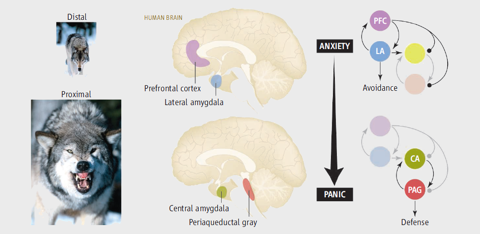 * Hypervigilance for Fear. 2012 / D. Terburg et al., Utrecht University, Utrecht, The Netherlands Human-neuroimaging data show that both the PAG and Central-Medial Amygdala (CMA) are activated when threats are imminent and unavoidable (panic, phasic fear). * Neural activity associated with monitoring the oscillating threat value of a tarantula. / 2010 fMRI study - 1Medical Research Council-Cognition and Brain Sciences Unit, Cambridge CB2 7EF, United Kingdom Increased experiences of fear coincided with augmented activity in a cascade of fear-related brain networks including the Periaqueductal Gray, Amygdala, and bed nucleus of the stria terminalis. Activity in the Amygdala was also associated with monitoring the tarantula's threat value as indexed by its direction of movement. * Amygdala activation during spontaneous Panic Attacks / 2011 - T.Dresler et al. Two anxiety disorder patients who spontaneously experienced a panic attack during an fMRI. * Amygdala activation to threat under attentional load / 2011 - T.Straube, Friedrich-Schiller-University, Germany Simultaneously increased amygdala and visual cortical activation to threat vs neutral pictures in phobic individuals, compared with controls, occurring regardless of attentional load. PANIC & AGORAPHOBIA ASSOCIATED WITH PTSD Figure 1: schematic of Amygdala, the brain site most critical for Fear Learning. Information regarding the conditioned stimulus (CS) and unconditioned stimulus (US) is transmitted to the amygdala by way of sensory areas in the thalamus and cortex. Within the amygdala, the critical plasticity underlying the acquisition of fear conditioning is thought to occur in the lateral amygdala and the lateral portion of the central nucleus (CEl). The medial division of the central nucleus of the amygdala (CEm) projects to various brain areas that produce fear and panic symptoms seen in people with fear-related disorders. LA, lateral nucleus; BLA, basolateral nucleus; ITC, intercalated cells. 
Figure 2: model for the development of fear-related disorders. Certain individuals are predisposed to the development of fear-related disorders on the basis of early life experience and genetic background, among other risk factors. When a traumatic event occurs, people learn to fear the cues that are associated with the traumatic event, and this memory consolidates over the course of the subsequent hours and days. The expression of fear comes in several different forms, including flashbacks of the traumatic event, nightmares, avoidance of situations that trigger memory for the traumatic event and altered sympathetic responses such as increased startle. Additionally, fear may generalize to cues not associated with the initial traumatic event in those people who go on to develop a fear-related disorder.  * Psychophisiology of Post-traumatic Stress Disorder / 2010 - Katie Boyts, L.M.T. ReturningVeterans.org PTSD is hyperactive Amygdala (react first, think later) [..] Remove amygdala, remove fear * Excitatory effect of corticotropin-releasing hormone on the acoustic startle reflex / 1997 - Patients with PTSD show evevated cerebrospinal-fluid concentrations of CRF which may sensitize (chronic activation) CRF systems in Amygdala and BNST. * PTSD sleep research, an update / 2010 - Steve Woodward, National Center for PTSD That downregulation of amygdala function can result in better sleep. In one study, rhesus macaques that had been chemically amygdalectomized exhibited both more sleep overall and a higher percentage of REM sleep than controls (Benca et al., 2000) OTHER STUDIES Synaptic plasticity in the basolateral amygdala complex (BLA) mediates the acquisition of associative memories, both positive and negative. Different populations of BLA neurons may encode fearful or rewarding associations. * Departments of Psychiatry and Psychology, University of Wisconsin / 2001 Khalin, Shelton, Davidson et al. Primate Amygdala mediates acute fear but not behavioral and physiological components of anxious temperament. * REM sleep de-potentiates amygdala activity to previous emotional experiences / 2011 - Els van der Helm, Justin Yao, Shubir Dutt, Vikram Rao, Jared M. Saletin, and Matthew P. Walker Specifically, the marked suppression of Central Amygdala (CeA) adrenergic neurotransmitters during REM coupled with activation in amygdala-hippocampal networks that encode salient events, is proposed to (re)process and de-potentiate previous affective experiences, decreasing their emotional intensity. [..] Here, we demonstrate that REM sleep physiology is associated with an overnight dissipation of Amygdala activity in response to previous emotional experiences, altering functional-connectivity and reducing next-day subjective emotionality. * Sistema enzimatico di destrutturazione delle reti proteiche nellAmigdala / 2009 In pratica, spiega in un commento su SCIENCE Tommaso Pizzorusso dell'Istituto di Neuroscienze del CNR di PISA, il cervello infantile è più plastico e, ancora privo di questa rete di materiale proteico, può dimenticare più facilmente un ricordo traumatico. Poi quando la rete si forma i ricordi di eventi negativi si fissano in modo indelebile. Ma sciogliendo i proteoglicani condroitin fosfato con un enzima ad hoc, Gogolla ha mostrato di poter rendere nuovamente più malleabile anche il cervello adulto e quindi di poter eseguire l'esercizio di rimozione del trauma. Questo studio apre dunque le porte a nuovi meccanismi potenzialmente applicabili su chi soffre di stress post-traumatico o fobie. * MRI-guided selective laser Amygdalohippocampectomy / 2013 - 67° AES neurosurgery congress, Whasington DC / Less collateral effects on amygdala and hippocampus removal with laser tecniques compared with traditional surgery. (medscape.com/viewarticle/817498) |
 CeA
CeA |
download all researches (pdf) not available at the moment.. |
share:
PRINT THIS PAGE
|
|
| AMYGDALA.LINK (C) 2015/2016 ARR - - - - - - Special THANKS to Paternita.info/Fatherhood and PubMed.gov |